Mysteries of the poisonous amphibians
Many brightly colored frogs and salamanders have enough toxins in their skin to kill multiple people. How do they survive their own noxious weapons?
Support sound science and smart stories
Help us make scientific knowledge accessible to all
Donate today
From the brightly colored poison frogs of South America to the prehistoric-looking newts of the Western US, the world is filled with beautiful, deadly amphibians. Just a few milligrams of the newt’s tetrodotoxin can be fatal, and some of those frogs make the most potent poisons found in nature.
In recent years, scientists have become increasingly interested in studying poisonous amphibians and are starting to unravel the mysteries they hold. How is it, for example, that the animals don’t poison themselves along with their would-be predators? And how exactly do the ones that ingest toxins in order to make themselves poisonous move those toxins from their stomachs to their skin?
Even the source of the poison is sometimes unclear. While some amphibians get their toxins from their diet, and many poisonous organisms get theirs from symbiotic bacteria living on their skin, still others may or may not make the toxins themselves — which has led scientists to rethink some classic hypotheses.
Deadly defenses
Over the long arc of evolution, animals have often turned to poisons as a means of defense. Unlike venoms — which are injected via fang, stinger, barb, or some other specialized structure for offensive or defensive purposes — poisons are generally defensive toxins a creature makes that must be ingested or absorbed before they take effect.
Amphibians tend to store their poisons in or on their skin, presumably to increase the likelihood that a potential predator is deterred or incapacitated before it can eat or grievously wound them. Many of their most powerful toxins — like tetrodotoxin, epibatidine and the bufotoxins originally found in toads — are poisons that interfere with proteins in cells, or mimic key signaling molecules, thus disrupting normal function.
That makes them highly effective deterrents against a wide range of predators, but it comes with a problem: The poisonous animals also have those susceptible proteins — so why don’t they get poisoned too?

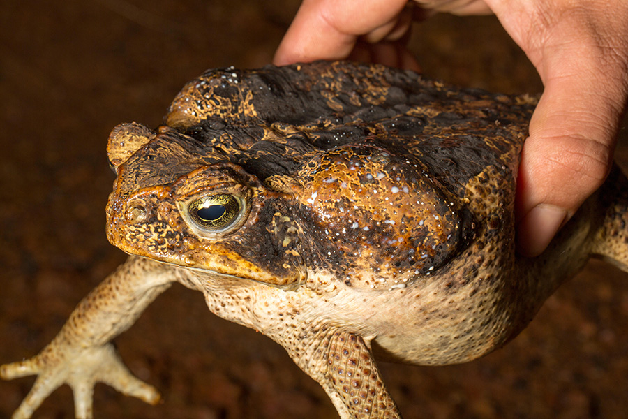
Toads such as this cane toad exude a toxin — the whitish blobs visible here — from glands behind the head. When researchers milk those glands to remove the toxin, the toads activate genes in toxin-related biosynthetic pathways, suggesting that the toads make the toxin themselves.
CREDIT: WWW.PQPICTURES.CO.UK / ALAMY STOCK PHOTO
It’s a question that evolutionary biologist Rebecca Tarvin took up when she was a graduate student at the University of Texas at Austin. Tarvin opted to study epibatidine, one of the most potent poisons of the thousand-plus known poison frog compounds. It’s found in frogs such as Anthony’s poison arrow frog (Epipedobates anthonyi), a small, ruddy creature with light-greenish-white splotches and stripes. Epibatidine binds to and activates a receptor for a nerve-signaling molecule called acetylcholine. This improper activation can cause seizures, paralysis and, eventually, death.
Tarvin hypothesized that the frogs, like some other poisonous animals, had evolved resistance to the toxin. She and her colleagues identified mutations in the genes for the acetylcholine receptor in three groups of poison frogs, then compared the activity of the receptor with and without the mutation in frog eggs. The mutations slightly changed the receptor’s shape, the team found, making epibatidine bind less effectively and limiting its neurotoxic effects.
That helps to solve one problem, but it presents another: The mutations would also prevent acetylcholine itself from binding effectively, which would disrupt normal nervous system functions. To address this second problem, Tarvin found, the three groups of frogs each have another mutation in the receptor protein that again changes the receptor’s shape in a way that allows acetylcholine to bind but still rejects epibatidine. “This is a series of very slight tweaks,” Tarvin says, which make the receptor less sensitive to epibatidine while still allowing acetylcholine to perform its usual neural duties.
Epibatidine, a potent toxin used by some poison frogs, works by binding to the same receptor as the neurotransmitter acetylcholine (left). This improperly activates the receptor, disrupting normal nerve activity. In response, the poison frogs have a mutation in their receptor that changes its shape so epibatidine no longer binds as effectively (center) — but neither does acetylcholine. So the frogs have evolved a second change in the receptor’s shape that restores acetylcholine’s ability to bind while still excluding epibatidine, re-establishing normal nerve function.
Tarvin, now at the University of California, Berkeley, is researching how animals evolve to cope with toxins, using a more tractable experimental organism, the fruit fly. To that end, she and her colleagues fed food containing toxic nicotine to two lineages of fruit flies that differed in their ability to break down nicotine.
When the researchers exposed fly larvae to predators — parasitic wasps that laid eggs in the flies — both groups of flies were protected by the nicotine they ate, which killed off some of the developing parasites. But only the faster-metabolizing flies benefited from their toxic diet, because the slower-metabolizing flies suffered more from nicotine poisoning themselves.
Tarvin and her students are now working on an experiment to see if they can induce the evolution of adaptations, such as those she identified in the frogs’ proteins, by exposing generations of flies to nicotine and wasps, then breeding the flies that survive.
Fishing for poisons
Poisonous animals must do more than survive their own toxins; many of them also need a way to safely transport them in their bodies to where they’re needed for protection. Poison frogs, for instance — which obtain their toxins from certain ants and mites in their diet — must ship the toxins from their gut to skin glands.
Aurora Alvarez-Buylla, a biology PhD student at Stanford University, has been trying to nail down which genes and proteins the frogs use for this shipping. To do so, Alvarez-Buylla and her colleagues used a small molecule she describes as a “fishing hook” to catch proteins that bind to a toxin — pumiliotoxin — that the frogs ingest. One end of the hook is shaped like pumiliotoxin, while the other end bears a fluorescent dye. When a protein that would normally bind to pumiliotoxin instead latches onto the similar hook, the dye allows the researchers to identify the protein.

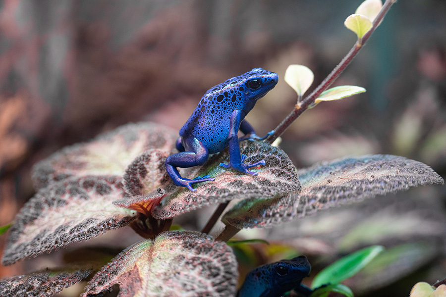
Poison frogs like this one get their toxins from animals in their diet. To find out how the frogs transport the poisons from their gut to their skin, scientists have gone on molecular fishing expeditions to see what binds to the toxin.
CREDIT: TIMO VOLZ / UNSPLASH
Alvarez-Buylla expected her hook to catch proteins similar to saxiphilin, which is thought to play a role in transporting toxins in frogs, or other proteins that transport vitamins. (Vitamins, like toxins, are usually scavenged from the diet and then moved around the body.) Instead, she and her fellow researchers found a new protein, similar to a human protein that transports the hormone cortisol. This new transporter, they found, can bind to multiple different toxic alkaloids found in different species of poison frogs. The similarity suggests that the frogs have borrowed the hormone-transporting system to also transport toxins, says Lauren O’Connell, Alvarez-Buylla’s PhD advisor at Stanford and a coauthor of the paper, which is still to be formally peer-reviewed.
This may explain why the frogs aren’t poisoned by the toxins, O’Connell says. Hormones often become active only when an enzyme cleaves their carrier, releasing the hormone into the bloodstream. Similarly, the new protein may bind to pumiliotoxin and other toxins and prevent them from coming into contact with parts of the frog nervous system where they could cause harm. Only when the toxins reach the right spot in the frogs’ skin would the toxin-carrying protein release them, into skin glands where they can be safely stored.
In future work, the scientists aim to understand exactly how the new protein can bind to several different types of toxins. Other known toxin-binding proteins, like saxiphilin, tend to bind tightly to just a single toxin. “What’s special about this protein is that it’s a little bit promiscuous in who it binds to, but also there’s some selectivity there,” says O’Connell. “How does that work?”
Turning toxic
While poison frogs definitively get their toxins from the food they eat, the source of toxins used by other poisonous amphibians is not always clear-cut. Amphibians such as toads, it appears, may make their own poisons.
To show this, TJ Firneno, an evolutionary biologist at the University of Denver, and his colleagues manually emptied the toxin glands of 10 species of toads by squeezing the glands (“It’s like popping a zit,” Firneno says, and is harmless to the toads), then looked at which genes were most active in those glands 48 hours later. The hypothesis, says Firneno, was that genes especially active after the glands are emptied could be involved in toxin synthesis.
Firneno and his colleagues identified several activated genes that are known to be part of metabolic pathways for creating molecules related to toxins in plants and insects. The genes they identified, Firneno says, can help point scientists in the right direction for further investigations into how toads may make their toxins.
Other amphibians may rely on symbiotic bacteria for their toxins. In the United States, newts of the genus Taricha are among the country’s most toxic animals. Though they look harmless, individual newts from some populations of these ancient creatures contain enough tetrodotoxin to kill numerous people. Many scientists believed the newts made the toxin themselves. But when a team of researchers collected bacteria from the newts’ skin, then cultured individual microbial strains, they found four types of tetrodotoxin-producing bacteria on the amphibians’ skin. That’s similar to other tetrodotoxin-containing species, such as crabs and sea urchins, where scientists agree that bacteria are the source of the toxin.

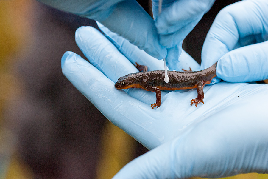
Newts in the genus Taricha, like this one, are among America’s most toxic animals. Scientists are still unsure whether the newts make deadly tetrodotoxin themselves or borrow it from bacteria living on their skin.
CREDIT: GEOFFREY GILLER
The origin of the toxin in these newts has broader ramifications, because they — and the garter snakes that eat them — are poster animals for what has been considered a classic example of coevolution. The snakes’ ability to eat the highly toxic newts is evidence that they have coevolved with the newts, gaining resistance so that they can continue to eat them, some scientists think. Meanwhile, the newts, the idea goes, have been evolving ever-greater toxicity to try and keep the snakes at bay. Scientists refer to this kind of escalating competition as an evolutionary arms race.
But in order for the newts to participate in such an arms race, they have to have genetic control of the amount of toxin they produce so that natural selection can act, says Gary Bucciarelli, an ecologist and evolutionary biologist at the University of California, Davis, who coauthored a re-evaluation of the arms race idea in the 2022 Annual Review of Animal Biosciences. If the tetrodotoxin actually comes from bacteria on the newts’ skin, it’s harder to see how the newts could turn up the toxicity. The newts could conceivably coerce the bacteria to pump out more tetrodotoxin, Bucciarelli says, but there’s no evidence that this happens. “It’s certainly not this very tightly linked, antagonistic relationship between newts and garter snakes,” he says.
Indeed, at the field sites where Bucciarelli works in California, he’s never actually witnessed a garter snake eating a newt. “If you follow the literature, you’d think that there are snakes just picking off newts like crazy at the edge of a stream or a pond. You just don’t see that,” he says. Instead, the snakes’ resistance to tetrodotoxin could have arisen for some other reason, or even by evolutionary happenstance, he says.
The newts’ toxin source is far from nailed down, though. “Just because you have bacteria that do something that live on your skin, doesn’t mean that’s the source in newts,” says biologist Edmund Brodie III, who was among the scientists that first put forward the arms race hypothesis between the snakes and newts more than 30 years ago. Brodie notes that other researchers have found that newts contain molecules that, based on their structures, may be part of a biological pathway for newts to synthesize their own tetrodotoxin. Still, Brodie says of the study showing that bacteria found on the newts can produce tetrodotoxin, “it’s the best thing we have so far.”
Brodie’s instinct is that one way or the other, the newts control their tetrodotoxin production, whether that’s by making the tetrodotoxin themselves or somehow manipulating their bacteria. The presence of bacteria as a third player in the newt-snake war would just make it an even more interesting system, he says.
Bacterial communities on the skin and in the glands of Taricha newts. Some of these bacteria, researchers have shown, are capable of producing tetrodotoxin. This suggests, but does not yet prove, that the newts may get their toxins from their skin bacteria.
One major barrier in determining whether the newts can make tetrodotoxin on their own is that no full genome has been published for Taricha newts. “They have one of the largest genomes of any animal we know of,” says Brodie.
Studying the ways that poison animals adapt and use toxins, just like much basic science research, has inherent interest for researchers who seek to understand the world around us. But as climate change and habitat destruction contribute to an ongoing loss of biodiversity that has hit amphibians especially hard, we’re losing species that not only have intrinsic importance as unique organisms but are also sources of potentially lifesaving and life-improving medicines, says Tarvin.
Epibatidine, tetrodotoxin and related compounds, for example, have been investigated as potential non-opioid painkillers when administered in tiny, controlled doses.
“We’re losing these chemicals,” Tarvin says. “You could call them endangered chemical diversity.”
10.1146/knowable-042423-2
TAKE A DEEPER DIVE | Explore Related Scholarly Articles


