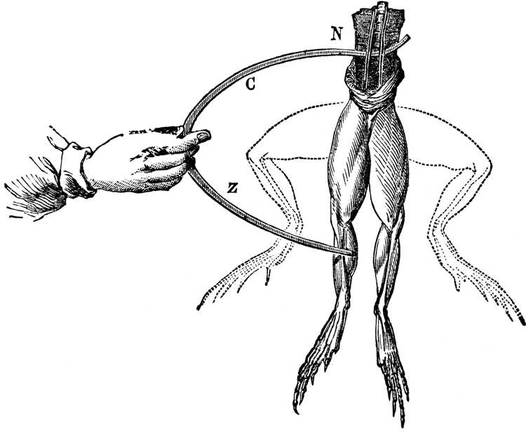
In the early nineteenth century, the nature of electricity remained a mystery to scientists. Experiments from the era showed that a spark could make dead frogs’ muscles twitch, or even set human corpses into convulsions — supernatural fodder that may have inspired Mary Shelley’s famed novel, Frankenstein. More than 200 years later, all the ways that electricity acts in the human body still aren’t completely understood. It is clear, however, that electric signals play a major role in the body’s early development.
Scientists like Michael Levin of Tufts University have discovered that cellular charges control how and where a structure forms in a developing embryo. Even more surprising, he’s found that it’s possible to manipulate bodily forms just by changing the voltage patterns of its cells.
Using this basic technique, Levin and his colleagues have successfully grown functioning third eyes on the backs of tadpoles. They’ve triggered brain damage in frog embryos by blocking key neural structures from forming — and then reversed the damage by changing the electrical charge of the developing brain cells. Although this work is still deeply experimental, Levin thinks it could have a major impact on the fields of medicine, biology and biochemistry. He imagines one day using bioelectricity to reverse birth defects in the womb, treat cancer or even grow new limbs on amputees.
Levin, director of the Allen Discovery Center at Tufts and coauthor of an article in the 2017 Annual Review of Biomedical Engineering on the topic, recently spoke with Knowable Magazine about the state of bioelectric research and his thoughts on its future prospects. This conversation has been edited for length and clarity.
In the context of biology, what does an “electrical signal” really mean?
Well, in the membrane that surrounds every cell, there are embedded proteins that can move ions — charged atoms — in and out of the cell. Things like potassium, chloride, sodium, protons, and so on. And inevitably, if you add more charged ions to one side of a membrane, you’ll generate an electrical potential across that cell surface. That’s basically what happens in a battery, where one side of the battery has a different amount of charge than the other.
It turns out that cells can actually use those charges to communicate. These signals are much slower-acting than impulses we’re used to hearing about in the nervous system — there, you’re talking about millisecond time scales for information flow, but in developmental bioelectricity, you’re talking about minutes or even hours. But ultimately, the electrical potential between cells can determine how certain tissues or structures develop.
How exactly do these electrical signals affect development in the body?
Bioelectric signals serve as a kind of a high-level master regulator switch. Their spatial distribution across tissues and intensity tells a region on an embryo, OK, you’re going to be an eye, or you’re going to be a brain of a particular size, or you’re going to be a limb, or you’re going to the left side of the body, that kind of thing.

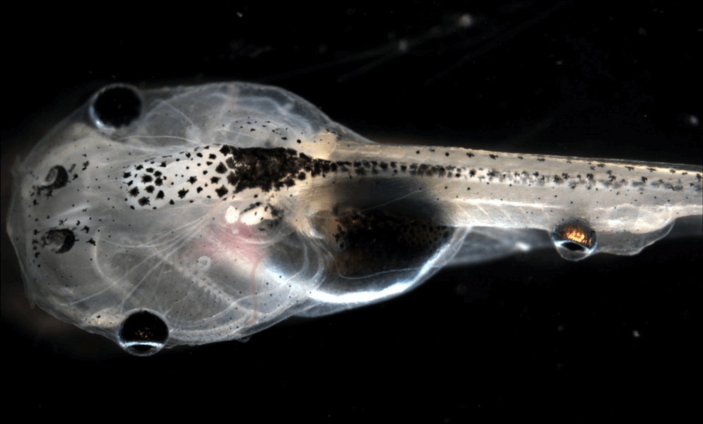
The sphere on the tail of this tadpole is actually a developing frog eye. By exposing the implanted tissue to certain neurotransmitter drugs, scientists were able to coax nerve tissue to grow from it. This successfully connected to the developing tadpole’s spinal cord, sending visual information to the brain and letting the otherwise blind tadpole see.
CREDIT: ALLEN DISCOVERY CENTER, TUFTS UNIVERSITY
You can actually see them forming in frog embryos. For example, electrically sensitive dyes reveal a pattern that we call the “electric face” — electrical gradients across the tissue that lay out where all the parts of the face are going to form later. It’s like a subtle scaffold for the major features of the anatomy, while a lot of the local details seem to be filled in by other processes that may or may not involve bioelectricity. If you change those electric signals in a developing embryo, it can have a major effect on how and where its structures form.
Can you give an example of how that works on a specific organ?
Sure. One of the things we wanted to study a few years back is how transplanted cells and tissues will develop in a foreign environment. We took the early eye structure from one frog embryo, and implanted it onto another embryo’s back. We were interested in two things: First, would the recipient be able to see out of that implanted eye on its back? Is the brain plastic enough to be able to actually see out of it? Second, we wanted to know, what is this eye structure going to do without a brain nearby? Where is it going to connect, and what are the neurons going to do?
What we discovered is that when you implant that structure in a developing tadpole’s back, the eye cells make a functional retina and optic nerve that sort of meanders around and tries to connect up in the spinal cord somewhere. But if you lower the electrical potential of the cells surrounding the implant, the eye structure goes crazy, and makes huge numbers of new nerves that emerge from it.
It turns out that emerging neurons can read the electrical signals of the tissue that they are sitting on. If the cells in that tissue have a polarized resting potential — meaning that they’ve accumulated negative charges inside each cell — the implanted eye forms an optic nerve and that’s the end of it. But if they’re depolarized, or have a lower charge, that gives the neurons a signal to overgrow in a very profound way. So we think this is an example of cells reading the electrical topography of their environment, and making growth decisions based on that information.
When sliced in half, a flatworm can normally regrow missing parts of its body. By manipulating its cells’ electrical charge, however, scientists can control which of these parts regenerate. By blocking the normal influx and outflux of charged ions from the flatworm’s cells, they can create a hyperpolarized state in both sides of the regenerating tissue, which prompts the worm to grow two tails. Or, they can create a depolarized state, leading to the formation of a second head to replace its amputated tail.
So if you change the bioelectric signals around the eye implant, it grows into the tadpole’s nervous system?
Yes. Not only is it growing into a complete eye structure, but it’s also functional. If you remove the tadpole’s existing eyes, the implant lets the otherwise blind animals see colors and moving shapes. In our study, we put blinded tadpoles in a shallow dish on top of an LCD monitor, and chased them around with little black triangles. The tadpoles consistently swam in response to the triangles' motion. We’re not able to tell if they have the same visual acuity as normal tadpoles, but they can definitely see out of that new implanted eye.

Active in the mid-eighteenth century, Luigi Galvani did seminal experiments on how electric signals activated muscles in the body — making the legs of a dead frog twitch after zapping them with electrodes (shown) — and was among the first scientists to discover bioelectricity.
CREDIT: LUIGI GALVANI / WIKIMEDIA COMMONS
How do you go about manipulating the electrical state of the cell or tissues?
We can do it with drugs that target ion channels in cells. Right now, something like 20 percent of all drugs out there are ion-channel drugs, things people take for epilepsy and other diseases, so they’re not hard to find. In our lab, we’re specifically making drug cocktails that target specific regions of the body. If you wanted to target the voltage of skin, for example, we might use a drug that opens or closes ion channels expressed solely in skin cells. You tune the cocktail of drugs to cause different reactions in different parts of the body.
You started in this field as a computer scientist. Do you see parallels between coding for a computer and tweaking electric signals in a biological setting?
Absolutely. On a fundamental level, I care about the information processing and algorithms in a system. It doesn’t matter if that system is made of silicon or living cells. To my mind, I’m a computer scientist, but I’m studying computation and information processing in living media.
People who have a computer science background understand that what’s fundamental about information sciences is not the computer itself — it’s the way it makes computations. Lots of different architectures and very distinct kinds of processes can be used to carry out a computation. People have made computers out of weird liquids, slime molds, even ants. So I think one of the most important things that computer science could teach the field of biology is this distinction between software and hardware.
Michael Levin’s colleague Dany Adams, who discovered what's called the electric face, created this time-lapse video that reveals how bioelectric signals help direct the construction of facial features in developing frog embryos (Xenopus laevis). Using fluorescent dyes that mark electrical potential, the bright cells are hyperpolarized (more negatively charged) than their dimmer neighbors.
In biology and chemistry, a body’s “hardware” — the cells and molecules inside it — is everything. But we’ve got to wrap our heads around the fact that these special kinds of hardware can in fact run many different kinds of software.
What do you mean by “software” in a biological sense?
The “software” in this case is the decisions of how cells cooperate to make a certain structure or tissue. That can be changed. You can take flatworms with one head, and by briefly altering electrical signals in their cells, get them to remember a new pattern that has two heads. Despite the fact that you have the same worm cells, you get a different outcome. And that kind of distinction between software and hardware is going to be really crucial as we tackle big issues of regenerative medicine and synthetic biology in the future.
What applications could this have in the medical world?
I think about that a lot. The most obvious ones are things like fixing birth defects. If we can understand and manipulate bioelectric signaling, we could potentially repair things that go wrong as an embryo forms. That’s one. We’ve actually induced some birth defects on animal embryos in the lab — and repaired them — by changing the electrical potential of certain cells.
Another one is fighting cancer. There’s a fair amount of research being done now on bioelectric signals as both a cause and a potential suppressor of cancer cells. You can normalize certain tumors by exposing them to specific drugs that change their electrical potential. Depending on the compounds you use, you can selectively affect only certain types of cells, like the ones in a tumor, while leaving the surrounding tissue intact. That’s pretty much ready for testing in mouse models.
A third area is regenerative medicine. If we can use electrical signaling to convince tissues and organs to grow after injury, we could replace entire structures or organs for patients. Bioelectricity gives you a great new set of control knobs with which to regulate cell behavior. It'll be much easier to build biological structures to suit once we understand these large-scale regulators like electrical signaling.
Editor's Note: This article was updated 8/10/18 to note Levin's role as director of the Allen Discovery Center at Tufts and to fix a typo in the description of ions in the cell. The description of the way the tadpoles swam in response to black triangles on an LCD screen was also clarified.


